
March 2020 Issue
Researcher Video Profiles
Yutaka Kano Professor, Department of Engineering Science, University of Electro-Communications
Molecular mechanisms controlling muscle contraction in the body
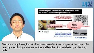
Muscle contraction is controlled by changes in various molecules in the body. In cells, the contraction and relaxation of calcium ions in a short time are regulated. To-date, many biological studies have revealed the changes at the molecular level by morphological observation and biochemical analysis by collecting cells.
However, these methods lack spatial and temporal information in the living body, that is, information on the dynamics in the living body. Yutaka Kano is working on bioimaging research to visualize in vivo molecules in living animal bodies. "To date, many fluorescent proteins and compounds have been made to visualize the inside of living organisms, but we have developed bioimaging technology to introduce these into living organisms," says Kano.
This video includes visualizations of the dynamics of calcium ions in muscle cells and examples of research using this observation method.
In skeletal muscle, there is a channel called TRPV1, which senses heat and allows calcium ions to flow into cells. In the video the fluorescence image shows that the more reddish the color, the higher the intracellular calcium ion concentration. In the middle heat-loaded group, heat increased the intracellular calcium ion concentration by heat, and inhibition of TRPV1 inhibited the increase in intracellular calcium ion concentration by heat.
"Hence, in this study, we succeeded in visualizing the dynamics of intracellular calcium ion concentration under thermal stimulation in an vivo environment," says Ryo Ikegami a graduate student in the Kano Laboratory.
Ayaka Tabuchi, a grad-student in the Kano Lab also appears in the video.
"I am focused on the change in intracellular calcium ion concentration due to exercise," says Tabuchi. "Changes in intracellular calcium ion concentration during exercise can be visualized by in vivo bioimaging."
In this study, the Kano Lab clarified the pathway of calcium ion influx into cells by exercise and the existence of a mechanism that homogenizes the accumulated information on intracellular calcium ion concentration in cells.
They are also working on visualization of vascular networks using two-photon microscopy and mitochondrial networks using photothermographic microscopy.
The goal is to elucidate physiological systems by applying the latest technology by developing fluorescent molecular probes and optical systems to bioimaging research and visualizing molecular dynamics in vivo.
Further information
Research Highlight: In vivo bioimaging: technology to elucidate sex-dependent differences in skeletal muscle function
Department website: http://blsc.xsrv.jp/en/