September 2016 Issue
Feature
Medical optical imaging: Innovative near infrared bioluminescent probes for deep tissue imaging
Shojiro Maki
Assistant Professor
Department of Engineering Science, Graduate School of Informatics and Engineering
Medical treatment of diseases: Transition from cure to replacement
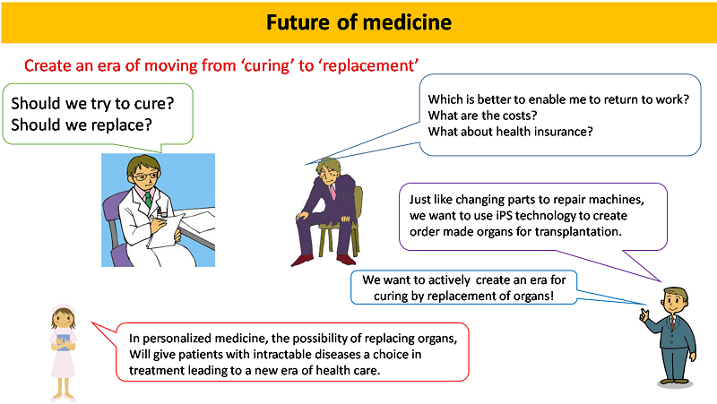
Advances in medical research have changed society through the ages. For example, the discovery and mass production of antibiotics revolutionized the treatment of tuberculosis and other diseases so-called incurable diseases. But what does the future hold in medical research and technology? Scientific reports indicate that advances in iPS research and related regenerative medicine will change our approach to treatment of intractable diseases such as ALS, and other global challenges including cancer, Ebola, SARS, and infectious diseases such as avian influenza.
"In the 21st century medical treatment will change from medical cure to medical replacement," says Shojiro Maki. "Regenerative medicine will enable the replacement of organs with congenital abnormalities. For example, type I diabetes will be cured by combining order-made personalized medicine with regenerative medicine. I want to contribute to this future by developing high precision 'optical in-vivo imaging technology' using for visualization of deep parts of the body far from the skin."
Optical in-vivo imaging technology
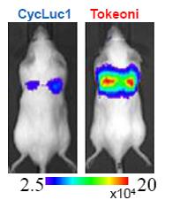
Conventional in-vivo optical imaging employs optical probes made of natural firefly bioluminescence with wavelengths of about 560 nm. The major problem with such probes is that this wavelength of light is mostly absorbed by biological materials (in particular blood) and does not reach the outside of the body where is could be detected with CCD cameras.
To overcome these shortcomings, Maki and colleagues used synthetic organic chemistry to produce firefly bioluminescence substances artificially, and importantly, they extended the wavelength to about 675 nm with their luciferin analogue called AlaLumine-HCL (Tokeoni). This wavelength yielded a major improvement of the in vivo transmission of 675 nm light, enabling the measurement of cancer cells metastasized to the lungs in mice, which was not possible with conventional light probes.
"Our 675 nm probes are a major advance in medical research," says Maki. "This material is AkaLumine (Wako Pure Chemical Industries, Ltd.) and is commercially available from Tokeoni (Sigma-Aldrich Co. LLC.). See Fig.1.
Future in vivo imaging technology
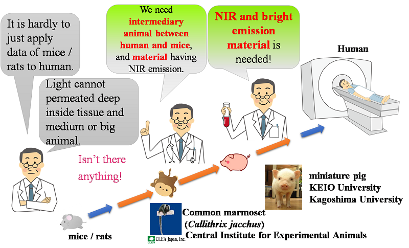
Maki and colleagues want to use this 675 nm imaging technology for basic research on regenerative medicine. "For example, we could create a new human liver using iPS technology," explains Maki. "The nurture it in the body of a pig, and when it is of the appropriate size, return it to a human. I envisage demand for this type of medical transplantation." Maki adds that realization of this technology would enable liver and heart transplantation to be performed in Japan without having to go overseas, and personalized medicine would reduce the amount immunosuppressive drugs used.
To achieve these goals it is necessary to develop high precision in-vivo imaging technology. Specifically, it is necessary to use high luminescent long wavelength light-emitting substrate and an appropriate optical enzyme (luciferase) both specially designed for the organs of pigs.
"We have already produced promising near infrared luciferin analogues," says Maki. "And patents for the appropriate light-emitting enzymes have been filed by collaborators at RIKEN (Japanese Patent Application No. 2016-165053). But the problem we have now is that the sensitivity of the existing equipment to long wavelengths is low, and the pigs cannot enter the measuring chamber."
Currently, we are planning a kick off meeting to launch a new 'pig optical imaging project' in collaboration with experts with in depth knowledge of conducting experiments with pigs, (Shizuoka Prefecture, small and medium-sized livestock research center) and staff at a measurement instruments company (Berthold Japan Ltd).
Publications
-
"A luciferin analogue generating near-infrared bioluminescence achieves highly sensitive deep-tissue imaging", Nature Communications 7, 11856 (2016), Doi: 10.1038/ncomms11856,
http://www.nature.com/articles/ncomms11856#abstract
Authors and affiliations
Takahiro Kuchimaru, Shun Mitsumata, Tetsuya Kadonosono & Shinae Kizaka-Kondoh: School of Life Science and Technology, Tokyo Institute of Technology, 4259, Nagatsuta, Midori-ku, Yokohama 226-8501, Japan.
Satoshi Iwano, Masahiro Kiyama, Haruki Niwa & Shojiro Maki: Graduate School of Informatics and Engineering, The University of Electro-Communications, 1-5-1 Chofugaoka, Chofu, Tokyo 182-8585, Japan. -
"Multicolor bioluminescence obtained using firefly luciferin", Current Topics in Medicinal Chemistry, 16 (24), 2648-2655, DOI: 10.2174/1568026616666160413135055,
http://benthamscience.com/journals/current-topics-in-medicinal-chemistry/volume/16/issue/24/,
Masahiro Kiyama, Ryohei Saito, Satoshi Iwano, Rika Obata, Haruki Niwa and Shojiro A Maki: The University of Electro-Communications. -
"Development of simple firefly luciferin analogs emitting blue, green, red, and near-infrared biological window light",
Tetrahedron, 69, 3847-3856, DOI: 10.1016/j.tet.2013.03.050,
http://www.sciencedirect.com/science/article/pii/S0040402013004134,
Satoshi Iwano, Rika Obata, Chihiro Miura, Masahiro Kiyama, Kazutoshi Hama, Mitsuhiro Nakamura, Yoshiharu Amano, Satoshi Kojima, Takashi Hirano, Shojiro Maki, Haruki Niwa: The University of Electro-Communications.
Further information
Shojiro MAKI Laboratory
http://www.uec.ac.jp/eng/research/introduction/opal-ring/0000470.html